
|
|
|
Clin. Cardiol. 23, 257 (2000)
This section edited by Edward A. Geiser, M.D.
Howard K. Baik, M.D., and Matthew J. Budoff, M.D., FACC
Harbor-UCLA Medical Center, Saint John's Cardiovascular Research Center, Torrance, California, USA
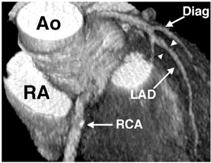
A 69-year-old male with history of systemic hypertension who was initially referred for the evaluation of chest pain underwent electron beam tomographic angiography. He was found to have a nonobstructing lesion in the mid left anterior descending (LAD) artery with 35% stenosis (Fig. 1). Subsequently, the patient continued to have atypical symptoms and was referred for catheterization angiography, which also demonstrated mid LAD lesion with 30% stenosis as measured with caliper (Fig. 2). The patient was continued on his antihypertensive medications and is currently being followed in clinic. He is asymptomatic on medical management. Adequate visualization of the coronary arteries is important when evaluating a patient for coronary artery disease. Thus far, catheterization angiography has been the standard of reference in evaluating coronary anatomy. Electron beam tomography (EBT) enables rapid acquisition of cardiac images, thus minimizing motion artifact, and allows visualization of cardiac anatomy with excellent spatial and contrast resolution. Given the noninvasive nature of the procedure, EBT is an attractive alternative means of assessing coronary anatomy in selected cases.
 |
|
| Fig. 1 Three-dimensional view using a volume rendering protocol from electron beam tomographic images demonstrating mid left anterior descending (LAD) stenosis (arrowheads). Also note the nonobstructive calcified lesion in the proximal right coronary artery (RCA arrow). LA, left atrium; Ao, ascending aorta; Diag, diagonal branch of the coronary artery. | Fig. 2 Left anterior oblique coronary angiogram during cardiac catheterization demonstrating mid LAD stenosis (arrowheads). |
Reference